Implant dentistry
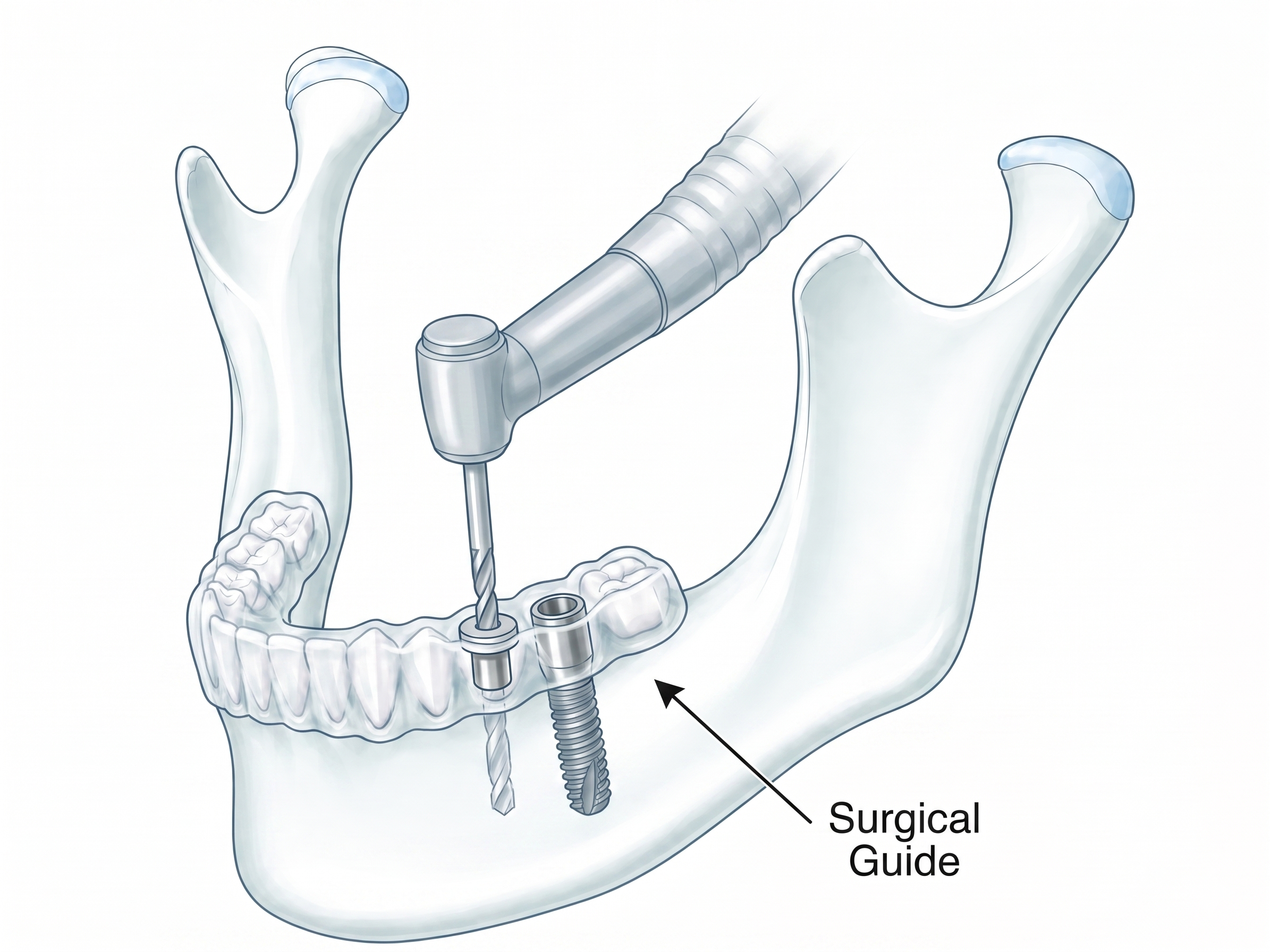
Implant drilling guide
An implant drilling guide transfers the planned implant axis into surgery. The guide is designed from the dental scan and radiological anatomy so that the sleeve position follows the prosthetic target while respecting bone volume and anatomical limits.
The design can include tooth supported seating, drilling sleeves, inspection windows, and support surfaces for a predictable intraoperative position.