Resection and immediate reconstruction

Tumor
Reconstruction

Virtual resection planning and fibula based mandibular reconstruction, from defect geometry to segment position, guide design, and final contour restoration.

Before you submit a case

Imaging & data requirements

How to acquire the CT/CBCT, dental and fibula donor imaging we need for accurate resection and reconstruction planning — our requirements from your side, on one printable sheet.

Download PDF
01 · The procedure

What Tumor Reconstruction Planning Does

When a tumour is removed from the jaw, the missing bone is rebuilt, most often using a section of the patient's own fibula (the slender bone of the lower leg). We plan the removal and the rebuild together in 3D before the operation.

The native jaw defines the gap to fill, the fibula provides the bone, and the new contour is checked against the bite, facial symmetry, and jawline before anything is made, so the surgical concept reaches the operating room with clear references.

Planning variables
Resection
planes matched to the surgical margin concept
Segments
fibula osteotomies adapted to the mandibular arc
Contour
inferior border and angle continuity restored
Fixation
plate and screw positions coordinated with segments
Transfer
cutting guides and reconstruction references documented
02 · Fibula reconstruction

Mandibular Defect to Fibula Based Reconstruction

The reconstruction is planned from both sides of the problem. The mandibular defect defines the target shape, while the fibula is divided into segments that can reproduce the lower border, angle, and ramus contour.

Fibula free flap

Segmented mandibular reconstruction

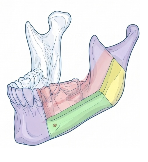
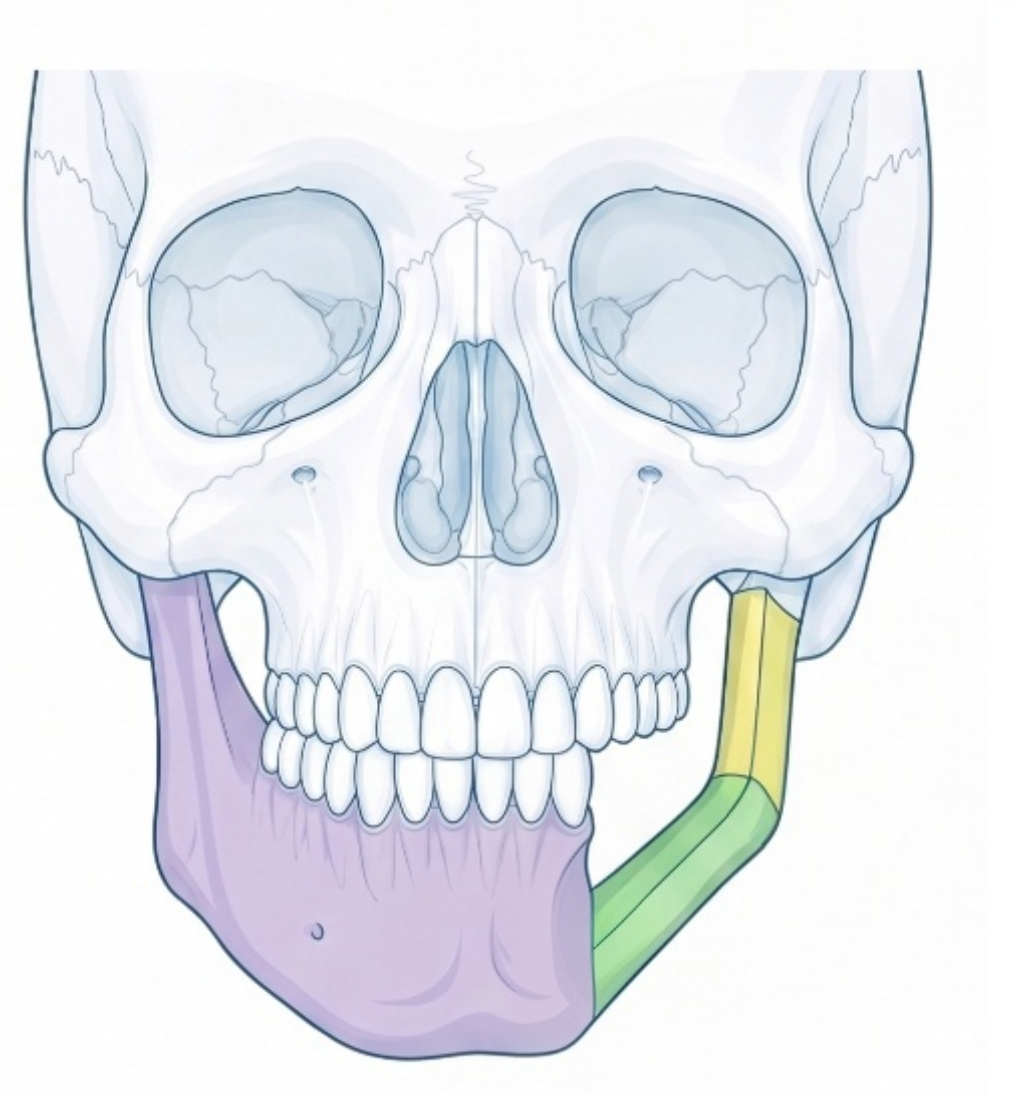
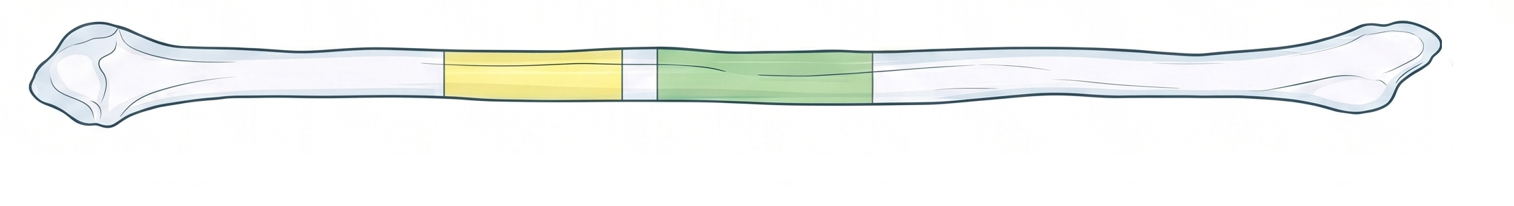
The fibula is virtually osteotomized into planned segments and arranged to match the mandibular defect. Segment length, angulation, and final position are controlled in relation to the residual mandible.

The design should make the reconstruction understandable before surgery: where to cut, how to rotate each segment, and how the final mandibular contour should be restored.

Segmental mandibular reconstruction with colored fibula segments
Segment position on the mandible
Frontal skull view with fibula mandibular reconstruction
Frontal contour restoration
Fibula donor bone divided into planned reconstruction segments
Fibula donor segment plan
03 · Surgical transfer

From Virtual Resection to Intraoperative Geometry

A reconstruction plan becomes useful when each digital decision is translated into a guide, a surface, a screw corridor, or a sequence that can be repeated during surgery.

01

Defect definition

The mandibular defect and residual bone are evaluated so that resection planes and reconstruction endpoints are clear.

02

Donor segmentation

The fibula is divided into planned segments that reproduce the desired mandibular curvature.

03

Guide design

Separate cutting guides are made for the fibula and the mandible, plus a model of the planned jaw to check the final shape.

04

Plate planning

Fixation is coordinated with segment position, screw access, and the final reconstructed contour.

04 · Planning logic

Reconstruct the Mandible Around the Planned Contour

The final reconstruction is not only a bone transfer. It is a controlled geometric transfer from the virtual plan to the patient, with the occlusal plane, inferior border, ramus height, and facial symmetry kept in view.

Shaped to the jaw

The fibula is divided into pieces that recreate the natural curve of the jaw.

Guided cuts

Cutting guides seat on stable bone, so each cut lands in the planned position.

Restored contour

The rebuilt lower border and jaw angle are checked for symmetry.

READY WHEN YOU ARE

Submit a case, receive a plan.

Send the patient's CT and dental scan through our secure channel. A planning proposal is typically returned within 2–5 working days, case-dependent.